


Fig. 5

Fig. 6

Fig. 7

Fig. 8

Fig. 9

Fig. 10
THE MECHANOBIOLOGICAL ACCOUNT OF VERTEBRATE LIMB DEVELOPMENT BY THE TENSILE ELASTIC FAILURE OF THE EMBRYONIC LIMB GIRDLES
Stuart Pivar, Mark McMenamin, and Peter Sheesley
ABSTRACT
This paper presents the discovery of a mechano-geometrical model accounting for the developmental formation of vertebrate limbs. The theorem demonstrates that limb development is the result of the elastic midline rupture failure of the limb girdles under tension caused by the enlargement of the underlying blastocoel.
The model demonstrates that the result of the application to failure of a force of tension in one direction upon an elastic orthogonal grid is a figure congruent with the archetypal vertebrate limb. The well known embryonic pectoral and pelvic girdles are composed of an elastic grid of cells.
KEY WORDS
Mechanobiology, blastula, gastrulation.
HISTORY
The study of limb development over centuries has amassed data serving the interests of anatomy in its widespread value in science and art. Although drawings of the steps of embryogenic development have existed since the seventeenth century, the accurate depiction of the steps of embryology were not perfected until the end of the nineteenth century by such noted workers as Wilhelm His Sr.
But the mechanism directing the complex ballet of embryonic cells that forms the organs remains a mystery. The embryologist is confronted with microscopic scale micron-thin, optically-indifferent membranes. Steps starting with limb buds are seen to seamlessly transform into hands and feet, much as an image appears on a photographic plate in a developing tray. Embryologists can literally make heads or tails of it, but little more.
In recent years, under the rubric of mechanobiology a sizeable segment of researchers have investigated non-genetic phenomena as candidate mechanisms of development. This paper presents a theory of limb development by non-genetic, mechanical means.
PREMISE
Limb development can be accounted for as the outcome of the deformation of predictable self-organizing geometrical patterns that occur in the stacking of cells produced by serial rounds of cell division.
Embryology describes development of the embryo as the deformation of the initial ball of cells, called the blastula. The structure of the blastula is unknown except for the appearance of two peripheral girdles–the pectoral and the pelvic–that originate the limbs.
Beginning with the premise that the blastula is in the form of a ball of an elastic tissue of rows and columns of cells self-organized in bands, the limbs are shown to be formed as two of the bands pull apart while the ventral midline bursts and separates. The dorsal recoil trajectory of the tense membrane of the two appendicular girdles causes the grid pattern to shrivel, forming the observed limb buds.
The steps of embryogenesis can be understood as though the blastula membrane is a thin elastic sheet stretched over a growing sphere. The parting of the ventral midline precipitates the elastic recoil of the two limb girdles, in resemblance of the deflating of a balloon. The predictable steps of this catastrophic snap-back are the reverse of the observed steps of vertebrate limb development.
COMMENTARY
Epigenesis is the sole mechanism guiding all-natural events, excepting those guided by human intelligence, which it can be argued is ultimately epigenetic as well.
The first three divisions of the fertilized egg cell are plainly visible to the microscope eye. The resulting eight cells of a theoretical cuboid are squashed together in their confining enveloping cortical sphere, in the embryological event called compression. Subsequent divisions create an optically incomprehensible drama that forms the ball of cells, called the blastula. Limb development begins when the pectoral and pelvic girdles each pull apart and separate at the ventral midline.
The first few divisions of the egg create simple forms that clearly occur by the mechanical forces on the cells, without any ostensible need for a guide. While the epigenetic stacking of cells can account for simple shapes, it is hard to conceive that an entire organism can be formed by the repeatable stacking of millions of cells. The claim of mechanobiology to generate the complex organism without the guidance of a genetic code is made plausible by the discovery of intercellular phenomena that can serve as clues, such as cell adhesion.
The gradual appearance of the image of a hand in the tissue of the embryological hand plate is like seeing an image appear on an exposed photo print sheet in the developing tray. To know the genes that trigger limb development is as useless as knowing the chemicals in the developing solution when it comes to understanding both kinds of development.
The presentation of the model is by a series of mechanical drawings depicting a hypothetical model of the unknown embryonic stages of limb development that are shown to accurately predict the musculoskeletal structure of the limb. The power of the algorithm to accurately predict nature surpasses coincidence.
The format of the presentation of the model is that of the Euclidean theorem where a hypothesis is proposed that can account for a phenomenon for which the cause is unknown. The phenomena of development are topological and geometrical. Data is graphic. This model is a developmental fate map which has no quantitative measurements and hence no quantitative data.
DEMONSTRATION
The mechanical account of limb development records the steps of the shriveling of the gridded pattern that composes the membrane of the pectoral and pelvic girdles. The topological deformations include:
The formation of the long bones by the axial rolling of a flat band of tissue, followed by the formation of split cuffs by axial compression;
The accordion failure of the limb axis by four or more bends;
The formation of claws by the 360 degree rotating trajectory of the digit tips that displace the symmetrical pattern of concentric circles at the tip to the locus of the paw-pads, fingerprints, and the extraneous prominences on limbs of tetrapods;
The appendicular musculoskeletal system in analogue with the accordion failure of a bilayer cylinder;
The accounting for the polymorphism in the vertebrate limb in the neotenic retardation or acceleration versus growth in ontogeny and phylogeny.
CONCLUSION
Limb development has for long offered a convenient laboratory model organism for developmental science. The presumed goal is to discover a system that can produce the form of the limb from axiomatic causes–the blueprint for the limb. This paper is the publication of the claim of primacy in the discovery. A corollary is that a code for the body in the genes is redundant.
REFERENCES
As the material presented is by definition unrelated to the state of the art of developmental science there is no continuum of theory.
Gould, S.J. Ontogeny and Phylogeny. Belknap Press of Harvard University Press, 2003.
Lewontin, R.C., It Ain’t Necessarily So: The Dream of the Human Genome and Other Illusions, New York Review of Books, New York, 2000.
Fields, C., and M. Levin. 2020. Does Evolution Have a Target Morphology? Organisms: Journal of Biological Sciences, vol. 4, no. 1, pp.57-76. DOI: 10.13133/2532-5876/16961.
Levin, M., and V. Bush. 2020. Reading and writing the morphogenetic code. Allen Discovery Center at Tufts University, Foundational White Paper.
Bessonov, N., M. Levin, N. Morozova, N. Reinberg, A. Tosenberger, V. Volpert. 2015. On a Model of Pattern Regeneration Based on Cell Memory. PLoS ONE 10(2): e0118091. https://doi.org/10.1371/journal.pone.0118091
Giaimo, C., 2020. When These Sea Anemones Eat, It Goes Straight to Their Arms. The New York Times. www.nytimes.com/2020/09/05/science/sea-anemones-arms.html.
. 2020. MRI scans of the brains of 130 mammals, including humans, indicate equal connectivity. Tel Aviv University. https://medicalxpress.com/news/2020-07-mri-scans-brains- mammals-humans.html
Serra, M., S. Streichan, M. Chuai, C. J. Weijer, L. Mahadevan. 2020. Dynamic morphoskeletons in development. Proceedings of the National Academy of Sciences. 117 (21) 11444-11449; DOI: 10.1073/pnas.1908803117
Sereno, M.I., et al. 2020. The human cerebellum has almost 80% of the surface area of the neocortex. PNAS. doi.org/10.1073/pnas.2002896117.
Wagner, D.S., K.R.W. Matthews. 2019. Human Embryo Research: What do we know and how do we know it? Rice University’s Baker Institute for Public Policy. https://www.bakerinstitute.org/media/files/files/fcd4841d/chb-pub-greenwall-her-022519.pdf
Bessonov, N., M. Levin, N. Morozova, N. Reinberg, A. Tosenberger, V. Volpert. 2015. On a Model of Pattern Regeneration Based on Cell Memory. PLoS ONE 10(2): e0118091. https://doi.org/10.1371/journal.pone.0118091
Burrows, L. 2020. Researchers use geometry and dynamics to better understand tissue organization. phys.org. https://phys.org/news/2020-05-geometry-dynamics-tissue.html
ILLUSTRATIONS
The copious illustrations have been accumulated over many years. The relative simplicity of the models makes detailed textual concordance redundant.
Figure 1
The overall limb developmental trajectory.
a, the pectoral and pelvic girdles in the blastula membrane
d, separation of the ventral midline
f, bone formation
g, axial accordion fold
Figure 2
a-m, limb development pattern
n-s, cross-section of neural tube formation
Figure 3
Bone formation
Figures 4, 5
1-11, the concurrent limb development and inversion as the inner cell mass
Figure 6
Schematic drawing of limb development
Figure 7
The origin of the musculoskeletal limb joint system in the compression accordion fold of the limb axis
Figure 8
Limb bone formation by axial warping and compression
Figure 9
Scapula and pelvis formation
Figure 10
Resume of vertebrate development
Figure 11
Formation of primate-form digits
Figure 12
Limb variants
Figure 13
The recoil phenomenon
Figure 14
The origin of fingerprints
Figure 15
Schematic analysis of digit development
Figure 16
Limb development polymorphism
Figure 17
Resume of limb development


TO REMOVE AFTER PUTTING IN ILLUSTRATIONS (BELOW)


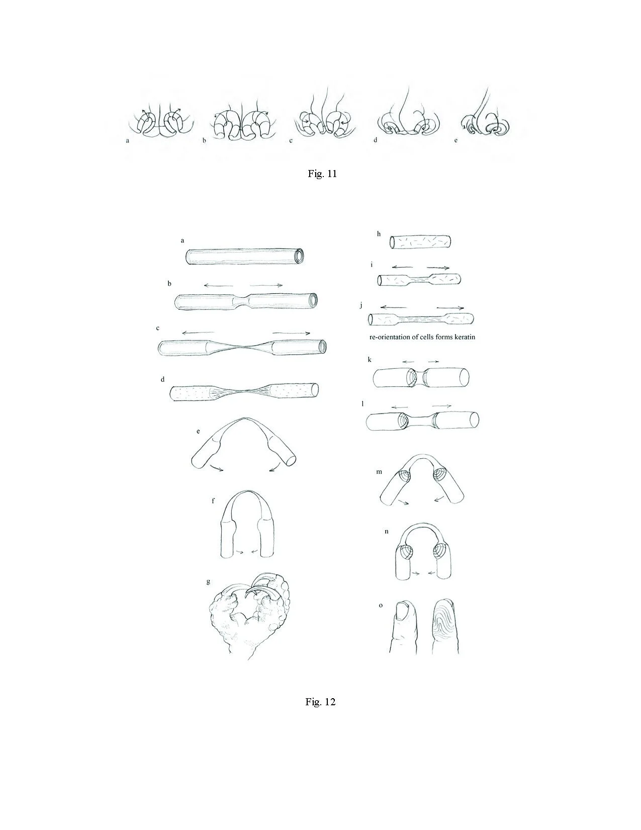

Fig. 1
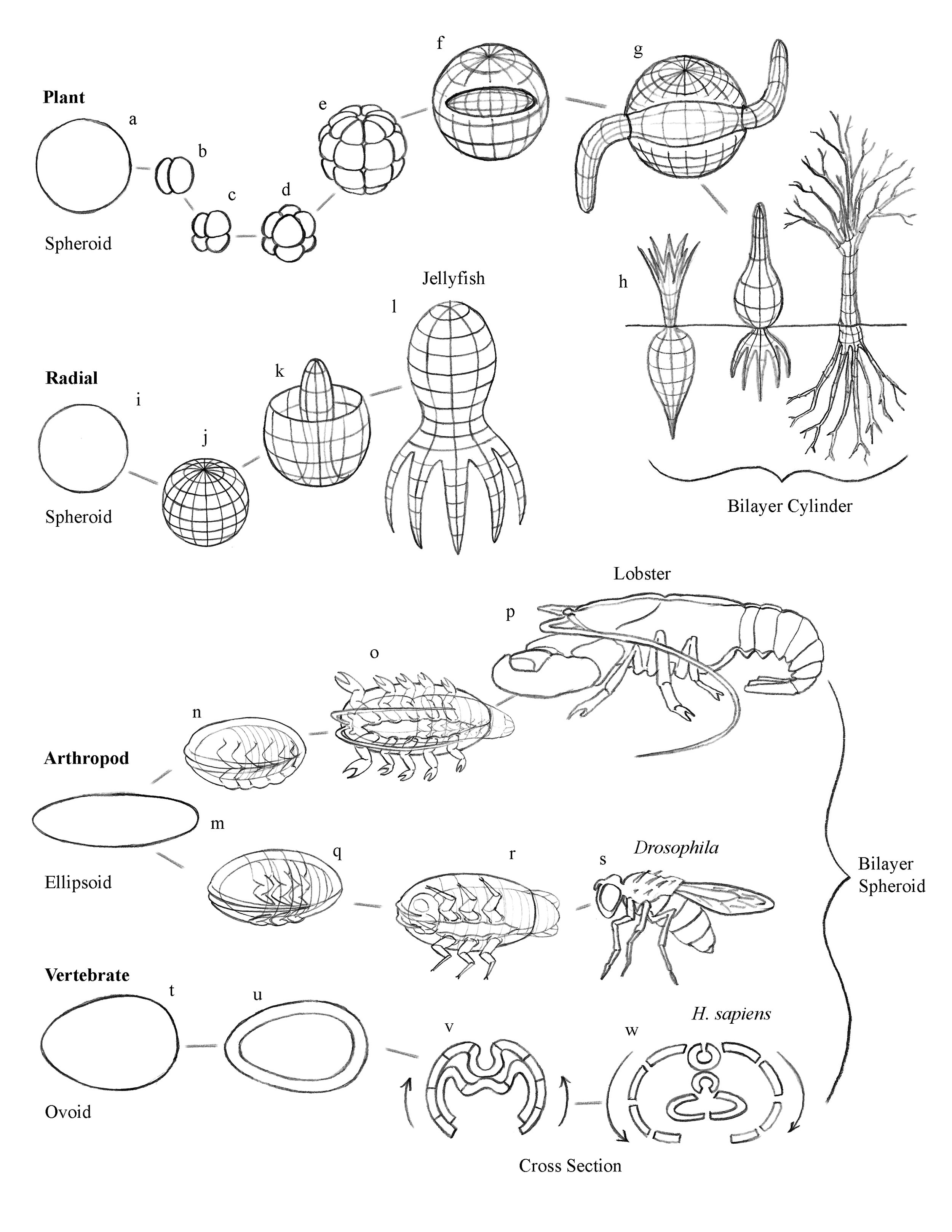
Fig. 2

Fig. 3
FIGURE LEGEND
Fig. 1 Flower morphogenesis from primordial germ plasm to flower and fruit. a, Germ plasm; b-d,
Axial extension; e-g, Roots/shoot differentiation; h-m, Flower/fruit development.
Fig. 2 Schematic drawings and photographs of model toroidal membrane. a-f, Schematic diverse
radial symmetry patterns of primordial germ plasm demonstrating thin axial tubes; g-i,
Photographs of vinyl toroidal membrane models.
Fig. 3 Ovary/fruit formation. a, Initial coaxial toroidal form; b-e, Withering of flower; f, Ovary/ fruit; g-i, Seed development; j, Generational reiteration.
Fig. 4 Schematic cross-section of primordial germ plasm membranes. a-l, Cross-section of various
fruits; m-r, Coaxial configuration of toroidal membranes; s-t, Schematic coaxial configura-
tion of membranes.
Fig. 5 Stem and branch morphology. a, Initial coaxial configuration of toroidal membranes; g-j, Extrusion of twig and branch forms; k, Flower meristem primordium.
Fig. 6 Origin of twig patterning. a-d, Invagination; e-f, Axial twist; g-i, Shoot; j, Twig/leaf pattern.
Fig. 7 Organismal model of the flowering plant. a-g, Shoot/root differentiation at opposite ends of
germ plasm; h-j, Leaf development; k-n, Flower development.
Fig. 8 Leaf morphology. a-d, Coaxial toroidal membranes; e-i, Extrusive growth of thorns and leaves; j-s, Leaf development.


